What to expect if you (or a loved one) need a CT scan.
Medically reviewed by Dr Roger Henderson and based on a text by Dr Carl J Brandt
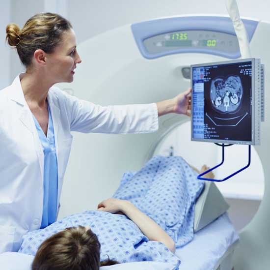
ACT (computerised tomography) scanner is a special type of X-ray machine. Instead of a single X-ray being sent through your body, like an ordinary X-ray, several beams are sent simultaneously from different angles.
This allows more detailed images from within the body to be constructed, which can then be analysed by a doctor. CT scanners may also be referred to as CAT scans (computerised axial tomography).
Unlike an MRI scan, where you are placed inside a tunnel, a CT scan comprises a doughnut-shaped machine, so you’re less likely to experience claustrophobia.
How does a CT scanner work?
X-rays from the beams are detected after they have passed through the body and their strength is measured. Beams that have passed through less dense tissue, such as the lungs, will be stronger, whereas beams that have passed through denser tissue, such as bone, will be weaker.
A computer can use this information to determine the relative density of the tissues examined. Each set of measurements made by the scanner is, in effect, a cross-section through the body which, once processed, is displayed as a two-dimensional picture on a monitor. Modern CT scanners can even produce three-dimensional images.
What are CT scans for?
CT scans are used for a variety of reasons, due to the fact CT imaging is one of the best and fastest ways of examining the chest, abdomen and pelvis, and because it can provide cross-sectional views and highly detailed images. It was originally designed to take pictures of the brain, but these days, it’s much more advanced and can be used to take pictures of virtually any part of the body.
CT scans allow doctors to inspect the inside of the body without having to operate or perform unpleasant examinations. It also allows surgeons to plan surgery prior to performing a procedure, and allows assessment of the results after a procedure has taken place.
Some of the most common uses of CT imaging include:
- Detecting different types of cancer(for example in the lung, bowel, liver and kidney)
- Examining patients with severe injuries
- Finding the cause of sudden rapid onset symptoms (such as breathlessness or abdominal pain).
- CT scan have also proven invaluable for cancer patients in pinpointing exactly where a tumour is, in preparation for radiotherapy.
- CT can detect and diagnose a number of vascular diseases, including abdominal aortic anueryms (AAAs), which occurs when the main artery running downwards in the abdomen becomes enlarged and prone to spontaneous rupture.
- It is also used to diagnose and analyse spinal and other skeletal problems and injuries, due to the fact it provides detailed images of even very small bones.
- Finally, the scanner is particularly good at testing for bleeding in the brain, aneurysms (when the wall of an artery swells), brain tumours and brain damage.
What happens before your CT scan?
If you need a CT scan, you will be asked about any recent illnesses or medical conditions, and whether you have a history of heart disease, asthma, diabetes, kidney disease or thyroid problems.
Prior to the procedure, you will likely be given a hospital gown to wear, and will be asked to remove metal objects, such as jewellery, glasses, dentures, hairpins, hearing aids and bras containing metal underwire. Where possible, piercings should also be removed.
If you are receiving an abdominal scan, you will be asked to refrain from eating for six hours before the test. You will then be given a drink containing gastrografin, an aniseed flavoured X-ray dye, 45 minutes before the procedure. This makes the intestines easier to see on the pictures. Sometimes, a liquid X-ray dye is injected into the veins during the test. This also makes it easier to see the organs, blood vessels or, for example, a tumour.
What happens during your CT scan?
A CT scan is a non-invasive, painless medical procedure. It usually takes approximately 10 to 30 minutes to perform, depending on the part of the body that’s being scanned, the number of pictures taken and the different angles required.
The scanner itself looks like a large doughnut, and the bed you lie on will pass through it. You will usually be asked to hold your breath during the scan. This is because any kind of motion, such as breathing or body movement, can affect the resulting image, making it harder to analyse and interpret.
When you’re ready to begin, the bed will move slowly backwards and forwards, to allow the scanner to take pictures of your body, although it will not touch you. During the scan, you will be alone in the exam room (unless there are special circumstances, for example if your child is the patient and you are accompanying them). During your scan, the technician will communicate with you via a speaker, and they will be able to hear and see you at all times.
Does a CT scan hurt?
The examination does not hurt, although some people find they become anxious lying within the scanner. Let your doctor and radiographer know if you feel panicky, or if you feel nervous of the ‘whirring’ noise the machine makes while working.
Is a CT scan dangerous?
The amount of radiation you will be exposed to is small, and therefore highly unlikely to cause you long-term harm. However, it’s important to be aware of the fact that a CT scan involves far more x-rays than a typical X-ray, so doctors only recommend them with good medical reason.
The risk is greatest to those who are pregnant, as radiation exposure can cause harm to the foetus – therefore CT scans are contraindicated in pregnant women, unless the benefits of performing the scan far outweigh the risks.
Risks are also greater in children than adults, so a CT is only recommended if a child has a serious condition that puts them at greater risk.
Some patients may experience side-effects due to allergic reactions to the liquid dye injected into the veins. In extremely rare cases, this dye has been known to damage already weakened kidneys. It’s important to let the X-ray doctors or technicians know if you have any allergies, asthma or kidney trouble, prior to having the X-ray dye injected.
Finding out your CT scan results
Your results will be analysed and interpreted by a radiologist – a doctor trained to supervise and interpret radiological procedures and images.
They will write a report based on your scan results, which will be sent either to your GP or to the doctor who originally referred you for the scan. Your doctor will then be in contact with you to discuss the results.
Net Doctor